Figure 3
Download original image
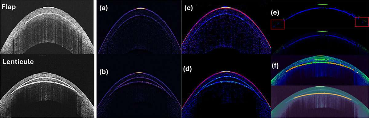
(Left) – Raw images of Figure 1. (Right) – The proposed image processing pipeline segments intrastromal lenticule and flap cuts by first enhancing the structures (a, b), then applying a segmentation algorithm to identify corneas (c, d) and consequently the domains of cuts (e, f). Panels in (e) illustrate the presence of sideband noise and its removal to achieve the correct flap geometry. Panel (f) demonstrates the optimization process for detecting the correct posterior (lenticule) cut (orange dots) using Algorithm 1.
Current usage metrics show cumulative count of Article Views (full-text article views including HTML views, PDF and ePub downloads, according to the available data) and Abstracts Views on Vision4Press platform.
Data correspond to usage on the plateform after 2015. The current usage metrics is available 48-96 hours after online publication and is updated daily on week days.
Initial download of the metrics may take a while.