Open Access
Figure 1
Download original image
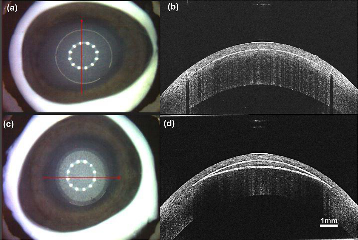
(a, b) Illustrate the aerial and OCT scan of the ex vivo intrastromal flap cut and (c, d) the lenticule cuts with SCHWIND ATOS on porcine eyes. Note the edge cut of the flap delineates as a circle in (a) and the reflection of plasma bubble layers (anterior and posterior) can be seen in (c).
Current usage metrics show cumulative count of Article Views (full-text article views including HTML views, PDF and ePub downloads, according to the available data) and Abstracts Views on Vision4Press platform.
Data correspond to usage on the plateform after 2015. The current usage metrics is available 48-96 hours after online publication and is updated daily on week days.
Initial download of the metrics may take a while.